Category

Uncategorized

ORPHANED WILDLIFE II

By Uncategorized No Comments

Orphaned Wildlife II

In our last article we discussed assisting orphaned or injured baby rabbits.  While Michigan is home to many different types of birds, such as water fowl, raptors, and more, the most common to live in urban areas are song birds.  You can reference our last article for general wildlife care guidelines and assistance on finding a licensed wildlife rehabilitator in your area.

In many cases baby song birds need just a little help or none at all.  There is a common myth that if a person handles a baby bird or nest, the parents will reject the baby, this is not true.  If you find a nestling, a young bird with few downy feathers, it can be placed back directly into the nest.  If the baby and the nest both fell out of the tree, but it is intact, you can place the baby back into the nest and put it back into the tree.

If the nest has been destroyed you can create a surrogate nest from a small box or plastic container.  Your new nest needs holes punched into the bottom for drainage, and lined with leaves, paper towels, or cloth.  Place the new nest in the tree, as high as you can safely manage, out of the sun and rain if possible.  Place the baby bird/s in the new nest and leave the area.  If parents do not visit the old or new nest, please contact a rehabilitator.

Fledglings are birds learning to fly, and in the process spend quite a bit of time on the ground as a result.  These little guys will be covered in feathers, like an adult but often have a few downy feathers.  The parents will often fly down to feed and protect the fledgling.  It is not uncommon for the parents to harass people or animals near the fledglings. If the fledgling is acting injured or sick, unable to flap its wings, bleeding, wings drooping unevenly, weak or shivering, please contact a rehabilitator.

While we are mainly discussing baby birds, window strikes by adult birds are also common.  If a bird strikes your window and is unable to fly away, place it in a paper bag or box with air holes.  Keep the bird in a safe, dark place for one to two hours.  The bird will need the help of rehabilitator if it is still unable to fly away within two hours.

Just like our baby rabbits, birds will need a safe container while waiting care of a rehabilitator.  Line the inside of a pet carrier, shoebox, or box with a soft cloth.  If using a box, don’t forget to add a few holes for ventilation.  Wearing gloves, gently transfer the bird to the prepared container.  Until the rehabilitators arrive, keep the bird warm by placing a heating pad under ½ of the box.  This allows the bird a place to cool down, if they become too warm.  If using a pet carrier, place a towel over the door to keep the light low.

To find a licensed wildlife rehabilitator, you can contact you local Humane Society, Audubon Society, local animal control, NRWA, and the International Wildlife Rehabilitation Council (IWRC).


http://www.nwrawildlife.org/

https://theiwrc.org/

ORPHANED WILDLIFE

By Uncategorized No Comments

Orphaned Wildlife

Each spring we see a new generation of young animals beginning to make their way in the world.  Some may still be under their parent’s care or just striking it out on their own.  Being young and naïve in the world, some individuals may find themselves in need of assistance.  The most common young animals found in residential areas, are birds and rabbits.  Today we will discuss rabbits and cover birds in the following article.

There are a few guidelines that must be addressed before we discuss caring for orphaned rabbits, and all wildlife in general.  These are wild animals and even young wildlife can be dangerous, safety is a priority.  Do not attempt to raise and/or care for injured wildlife.  The ultimate goal is to release wildlife back into the wild.  Special feeding and cages are used to allow injured wildlife to build up strength and practice hunting before their release.  Rehabilitators are licensed by the state and/or federal governments, not to mention they have spent many years learning about the animals they care for.  In many states it is illegal to possess a wild animal without a permit. Many wild animals have internal and external parasites that can be transmitted to humans.

Baby rabbits spend the majority of their time without their mother, who only visits the nest at dusk and dawn to protect the nest from predators.  It is very common for good Samaritans to think the rabbit nest has been abandoned or the babies are orphaned.   If a baby rabbit is found outside an intact nest you can place back in the nest and cover it with twigs and leaves.  Rabbits leave the nest when they are able to hop, about 5 inches long, their eyes and ears are open.  If you find a baby rabbit that is injured (bleeding, has puncture wounds, etc.), appears weak, thin, wrinkly baggy skin, or has been in a cat’s mouth, contact a rehabilitator.

If you need to intervene, the first step is to prepare a container for them.  Line the inside of a pet carrier, shoebox, or box with a soft cloth.  If using a box, don’t forget to add a few holes for ventilation.  Wearing gloves, gently transfer the rabbit to the prepared container.  Until the rehabilitators arrive, keep the rabbit warm by placing a heating pad under ½ of the box.  This allows the rabbit a place to cool down, if it becomes too warm.

To find a licensed wildlife rehabilitator, you can contact your local Humane Society, Audubon Society, local animal control,  NRWA, and the International Wildlife Rehabilitation Council (IWRC).

Vaping and Pets

By Safety, Uncategorized No Comments

VAPING AND PETS


What is an e-cigarette?

E-Cigarettes are electronic (battery operated) cigarettes. They come in all shapes and sizes. They can look like cigarettes, pens, pipes – they can be slender enough to fit into a wallet. “Vapes” work with an atomizer heating liquid containing nicotine, turning it into a vapor that is then inhaled. It creates a vapor cloud that looks like cigarette smoke.

How is this dangerous for my pet?

Nicotine has always been toxic to pets. Cases of toxicity are reported from cigarettes, nicotine patches, and nicotine gum. E-cigarettes carry new concerns if ingested by your pet because the nicotine is generally more concentrated than traditional nicotine products and the liquid form causes faster absorption. Liquid nicotine can be absorbed through the skin or mucous membranes of the mouth in dogs, cats, and humans. This causes a faster onset of symptoms. Additionally, the carrier may be propylene glycol and glycerin but ASPCA Poison Control states there have been reported cases of products containing diethylene glycol which can cause damage to the kidneys.

Pet-proof your home

Keep all of your vape supplies out of reach of your pets. Do not refill or change cartridges while your pet is nearby and never charge your e-cigarette in an area accessible to your pets. Dogs and cats are attracted to certain smells and cats have an intolerance to propylene glycol which is sometimes a carrier used in vaping liquids.

Symptoms of Nicotine Toxicity

Pets can show signs of toxicity very quickly, generally within 15 to 60 minutes of ingestion. Symptoms may include excitation or increased heart rate, abnormal breathing, salivating, vomiting, diarrhea, or agitation. More severe symptoms can include tremors, seizures, cardiac arrest and death.

If You Suspect Liquid Nicotine Ingestions

It is important you seek medical care immediately due to the rapid onset of symptoms. This is vital to your pets health. Call your veterinarian or an emergency veterinarian immediately.

What is Leptospirosis?

By Uncategorized No Comments

What is Leptospirosis?

Leptospirosis is a deadly bacterial disease of dogs and other mammals that affects the liver or kidneys. The bacteria (Leptospira) is found worldwide in soil and water.  It is commonly found in puddle water, lakes, and even wet grass.  Leptospira bacteria are commonly carried deer, skunks, raccoons, opossums, fox, rats and mice, but can be carried by almost any mammal, including people.  Leptospirosis is a zoonotic disease – meaning it can be spread between humans and animals.  According to the CDC, people can contract leptospirosis in the same ways your dog can – through contact with urine or other body fluids (except saliva) from an infected animal or by contact with other contaminated sources such as swimming in urine-contaminated water.

The diagnosis of Leptospirosis in dogs has been on the rise in Michigan.  According to the Michigan Department of Agriculture and Rural Development (MDARD), Reported Leptospirosis cases have doubled over the last six years.

The most common early signs of Leptospirosis in dogs are: decreased appetite, increase or decreased urine production, lethargy, vomiting, diarrhea, and abdominal discomfort.  Severely infected dogs show signs of lethargy, depression, loss of appetite, vomiting, fever, and increased thirst and urination. Dogs may develop jaundice and, in some cases, bleeding.  Illness typically develops quickly, sometimes in just a few days, and can be rapidly fatal. In comparison, dogs with mild infections may show little or no signs of illness and the disease may go undetected.

To vaccinate or not to vaccinate?  Well, Leptospirosis was very common forty years ago and thanks to vaccination, was under control.  However, it’s now considered a re-emergent disease.  This is due to different strains of Leptospirosis.  The older vaccine protecting against two serovars (a subdivision of species or subspecies distinguishable from other strains) but today’s second-generation vaccine provides protection against four serovars with fewer adverse reactions.


Pancreatitis

By Uncategorized No Comments

Pancreatitis

The center of our holiday celebration is large meals for our loved ones to share.  Our pets are so much a part of our family that we can’t help but to share with them too.  Unfortunately, our pets can get sick from eating human food.  This can range from GI upset including diarrhea and vomiting, to severe problems including intestinal blockages, bloat, and pancreatitis.  Pancreatitis (inflammation of the pancreas) can occur when a dog eats high fatty meals. It is commonly diagnosed over the holiday season.

The pancreas plays a large role in digestion.  It’s located next to the stomach and part of the small intestine.  In the GI system the pancreas secretes digestive enzymes to help break down food and neutralize stomach acid as food leaves the stomach.  The pancreas also produces insulin and glucagon which help regulate blood sugar levels in the blood stream.

Commonly, dogs with pancreatitis have a recent history of receiving table scraps or eating food from the garbage.  Cats can also suffer from pancreatitis however; it tends to be a chronic condition.  Other causes include metabolic disorders, tumors, toxins, certain medications, and trauma.  Pets that are obese are more susceptible, as are certain breeds such as Miniature Schnauzers, Miniature Poodles, and Siamese cats.

Pancreatitis is believed to be an activation of digestive enzymes within the pancreas causing inflammation and irritation.  Long term this can cause the pancreas to breakdown as the enzymes eat away the gland.  Eventually the digestive enzymes can make their way to the blood stream and affect other organs or cause an abscess (pocket of infection) on the pancreas itself.  Severe pancreatitis can be fatal, so treatment is vital.  Symptoms of pancreatitis include depression, poor appetite, vomiting, dehydration, and fever.  Some pets  also experience diarrhea and abdominal pain.

Treatment will vary based upon the severity of the disease and patient’s medical history.  Mild cases of pancreatitis can generally be treated on an outpatient basis with little or no long term damage to the pet.  Severe pancreatitis may require hospitalization, IV fluids, IV medications, and close monitoring.  Pancreatitis can develop into a long term, chronic condition.  Switching to low-fat diet and maintaining a healthy weight are recommended.

This holiday season avoid the temptation of feeding your pets’ table scraps.  Pets are such a part of our families that not sharing can lead to feelings of guilt.  Consider buying special treats, new brands of cookies, and a new toy or two this time of year.  If house guests cannot resist treating your dogs, have a dog safe cookie jar available.  With a little planning our pets can safely join in the food festivities without the worry of pancreatitis.

Veterinary General is located in Shelby Township, Michigan.  We offer traditional and alternative therapies such as Acupuncture, Chinese Herbals, and Cold Laser Therapy.  More information can be found at www.veterinarygeneral.com.             

Halloween Dangers

By Uncategorized No Comments

Autumn is full of color and full of fun.  The October schedule seems to be filled with harvest festivities  right up until the very last day, Halloween.  Generally a childhood favorite, Halloween is an evening of dressing in fun or scary costumes. But for pets, Halloween is not the “scary fun” that children enjoy – it can be very upsetting for our pets and a real challenge for us as pet owners.  There are many pet dangers associated with this “holiday”, including the obvious – Chocolate.

Because their theobromine metabolism process is significantly slower than that of a human, Chocolate is toxic for dogs.  Many people just don’t understand how dangerous it can be for their pet.  The greater the cocoa content, the greater the danger.  For example, Milk Chocolate has less cocoa than Semi-Sweet Chocolate which has less than Dark Chocolate.  Cocoa Powder is very concentrated and extremely dangerous to our pets.  Two ounces (2 oz) of Milk Chocolate ingestion may not be a toxic dose for a 40 pound dog but two ounces of Cocoa Powder could have a devastating outcome.   Keep all candy out of your pet’s reach.

Costumes are another concern.  Yes, it’s cute and may be fun to dress our pets but it’s important to realize when cute turns into scary for pets.  Costumes cause stress for many dogs and cats so limit the dress-up time unless you are certain your pet isn’t bothered by it.  And always be sure to check costumes for parts that could be choking hazards.

Additionally, when decorating, be sure your décor is not pet accessible – especially Jack-O-Lantern and  candles.  Decorations with a flame could be easily knocked over by our excited or anxious pets.

Finally, during trick-or-treating hours, kennel, contain, or keep pets safe in a separate room to avoid the stress of the excessive activity.  This will ease anxiety and keep them from running off, either with or away, from all those ghost and goblins.

Understanding Your Pet’s Blood Work

By Uncategorized No Comments

Understanding Your Pet’s Blood Work

This entry will examine routine blood work for pets.  Blood work can be described as your pet’s “internal examination.”  When you receive copies of your pet’s blood work the abbreviations and numbers can appear daunting.  Often we turn to the internet to research, but sometimes these searches lead to false or even frightening information.  In explaining blood work we will break it down into three categories the Complete Blood Count, organ chemistries, and electrolytes.

We will try to keep these explanations as simple as possible, and there is only so much one can do to make blood work sound fascinating to everyone.  Some things to also keep in mind while reading; this is not a list of every test we can run on a pet, it’s just the basics.  The abbreviations listed below may be slightly different depending on the laboratory used.

The CBC, Complete Blood Count

CBC’s are used to evaluate a pet’s blood, including platelets, red and white blood cells.  A CBC gives us information on hydration status, anemia, infection, the blood’s clotting ability, and the overall status of the body’s immune system.

  • HCT – Hematocrit measures the percentage of red blood cells in the blood. An abnormal result can indicate anemia or dehydration.
  • Hb and MCHC – Hemoglobin and Mean Corpuscular Hemoglobin Concentration measure the blood’s ability to carry oxygen throughout the body.
  • WBC – White Blood Cell counts measure the amount of immune system cells. Abnormal results can indicate disease, infection, or inflammation.  Individual types of white bloods cells are reported as a percentage then as an absolute number; which is the total number of cells in the blood.   Absolute counts are designated by “ABS” before or after the white blood cells’ name.  Types of white blood cells include:
    • NEUT SEGS – Segmented neutrophils are the most common white blood cell
    • BANDS – Band neutrophils are immature neutrophils
    • LYMPH – Lymphocytes
    • MONO – Monocytes
    • EOS – Eosinophils
    • BASO – Basophils
  • PLT – Platelets form blood clots to stop abnormal blood flowChemistry ProfilesChemistries evaluate organ ability to perform its “job” in the body.

Liver – The liver has many functions include filtering the blood as it leaves the small intestines and is responsible for taking substances such as medication then altering it to a form the body can use.

  • ALT – Alanine Aminotransferase can indicate liver disease, but doesn’t identify the cause
  • AST – Aspartate aminotransferase can indicate liver disease and muscle inflammation
  • ALKP – Alkaline phophatase can be elevated in patients with liver disease, Cushing’s disease, or in young patient with growing bones.
  • GGT – Gamma glutamyltranspeptidase can indicate liver disease or excess steroids
  • TBIL – Total Bilirubin can indicate liver disease or cause of anemia

Kidney – The kidneys most well known function is filtering the blood and excreting waste material in urine. The kidneys also secrete hormones that cause the bone marrow to produce and release red blood cells into the blood stream.

  • BUN – Blood Urea Nitrogen is an indicator of the kidney’s ability to filter and remove urea from the body. Urea is a waste product of protein metabolism.
  • CREA – Creatinine is another waste production that is filtered by the kidneys and can help determine the cause of an elevated BUN
  • PHOS – Elevated phosphorus is associated with kidney disease, hyperthyroidism, and bleeding disorders

Pancreas – The pancreas has two main functions one is the secretion of enzymes that help break down food the other is secreting insulin into the blood stream.

  • AMYL – Amylase breaks down starches and carbohydrates
  • LIP – Lipase breaks down fat

Other – These are chemistry results that covers multiple organs or don’t fit underneath any of the organs listed above.

  • ALB – Albumin evaluates hydration, intestinal, liver, and kidney disease.
  • CHOL – Cholesterol supports diagnosis of hypothyroidism, liver disease, Cushing’s Disease, and diabetes.
  • GLOB – Globulin can increase with chronic inflammation and certain diseases.
  • GLU – Glucose when high can be a sign of diabetes. When low it can cause seizures, collapse, or coma.
  • TP – Total protein is amount of protein found suspended in the liquid portion of blood and evaluates hydration status.

Chemistry Profiles

Electrolytes are important for maintaining fluid balances, blood clot formation, nervous and muscle system functions to name a few.

  • Ca – Calcium
  • Na – Sodium
  • K – Potassium
  • Cl – Chloride

When is Blood Work Requested

Blood work is used to diagnose, determine a cause of illness, monitor progress of an illness, and evaluate treatment.  Annual blood work on a healthy pet may seem like an odd request however; early detection gives you the best prognosis for treating disease.  An example is your pet’s liver and kidneys.  We can compare them to a rubber band that can be stretched for large tasks, small tasks, and are used repeatedly.  Like a rubber band, they have a remarkable reserve and use a small portion to perform their daily tasks, but can be stretched in times of need.  We never know when too much stress will cause the rubber band to snap.  If the organs are “stretched” any stress in the form of food changes, medication, or anesthesia may cause organ failure (the snap of the rubber band).

Evaluating Results

Blood work is a small piece of the puzzle that when combined with physical exam and history, allow the veterinarian to form a picture of the patient’s health and well-being.  Individual results above or below normal ranges may or may not affect the patient’s health.  An example is a slightly elevated BUN (kidney value) in a puppy presented for a routine spay at 6 months old.  Since the CREA (kidney value) is normal, the puppy is healthy upon physical examination, and eating a high protein puppy food the veterinarian isn’t concerned.  The elevated BUN is a byproduct of the puppy’s high protein diet being broken down in the body.


PET DENTISTRY

By Uncategorized No Comments

Spotlight On Dentistry: Can’t you just scrape that off his teeth?
February is Pet Dental Health Month and at Veterinary General we feel dental health is crucial to the overall health of your pet. Volumes of books can and have been written on dentistry but in this entry we are going to look at each step in the cleaning process and explain why they are important. Since advanced periodontal disease and treatment is beyond the scope of this article we will only look at the cleaning details of a patient undergoing a “Stage 1” (see Initial Evaluation below) dental cleaning.
Dental care is quickly evolving in veterinary medicine and old recommendations are being discarded in light of research. It is not uncommon to get questions about dental radiographs, pre-anesthetic blood work, home care, and why those items are even important in a younger dog with mild tartar. Cost is another concern, and we often have clients compare costs from previous pets or other practices. Through education we hope to share the value of the complete dental prophylaxis and show how it is not just simply “cleaning the teeth.”

Initial Evaluation
The dental therapy process begins with an oral examination of the pet. The veterinarian or veterinary technician will stage a pet’s teeth based upon amount of tartar and gum irritation visible. This visual inspection allows us to create and plan and estimated cost for the procedure. Pets with more severe periodontal disease require more advance treatments and are discussed on an individual basis.
Dental disease is “staged” from one through four, with four being most severe. Stage one dental disease is marked by halitosis (bad breath), mild calculus (tartar) build up, inflamed and red gum line. In stage two there is more calculus build up, the gum line will appear shiny, and swollen. Stage three is marked by swelling and recession of the gum line that may bleed with gentle probing. Stage four is identified by severe inflammation, pain, gum recession, bleeding, and purulent (pus) discharge. These are simply the visible signs, a more accurate assessment is made when dental radiographs and charting are completed (VetMed Team).

Blood Work and Anesthesia
Pre-anesthetic blood work is required for all patients receiving a dental cleaning. The liver and kidneys are responsible for converting drugs into a form the body can use then removing them from the body. If the liver and kidneys are not functioning properly the anesthetic protocol can be altered or the procedure aborted. Dental disease can cause changes in the rest of the body and blood work will help us identify any potential problems.
During anesthesia the patient is monitored by a licensed veterinary technician at all times. Monitoring includes heart rate, respiratory rate, temperature, percent of oxygen in the blood, and blood pressure. All patients have IV catheters placed, receive IV fluids during the procedure, and have endotracheal tubes placed in their trachea. IV fluids help maintain blood pressure during anesthesia and allow for injections to be given if needed. Endotracheal tubes have a small balloon on the end that can be inflated to prevent fluid from entering the lungs.

Dental Radiographs
The veterinary technician begins the dental process with radiographs. Radiographs are key component between a complete dental prophylaxis and just cleaning the teeth. Cleaning and observing abnormalities above the gum line ignores 60% of the tooth (Iams Partners for Health, 2003). Radiographs allow the veterinarian to evaluate the root of a tooth, bone loss around the root, and pockets (spaces between the gum and tooth root). If caries (cavities in people) is seen on the surface of the tooth a radiograph will allow the veterinarian to assess how far the lesion affects the tooth. After extractions an x-ray is often taking to make sure the entire root of a tooth has been extracted. Radiographs can also reveal roots of baby teeth that did not fall out, and adult teeth that never erupted through the gum line.

Charting
The first step with charting is a second oral exam. This allows the veterinary technician to note any abnormalities that were not observable on the patient while awake. Pictures are taken for both the patient’s records and the pet’s owners. The veterinary technician then begins to chart the oral cavity. They insert an instrument, called a probe, gently between the tooth and gums. A small depth is acceptable and requires no treatment however; bigger “pockets” need attention and indicate periodontal disease. The technician will also note any missing, fractured, discolored, or loose teeth.
Charting and dental radiographs are the foundation of the veterinarian’s treatment plan (VetMed Team). A treatment plan will include a diagnosis, recommendations for treatment (including extractions if needed), prognosis, and home care plan.

Dental Cleaning, Polishing, and Fluoride Treatment
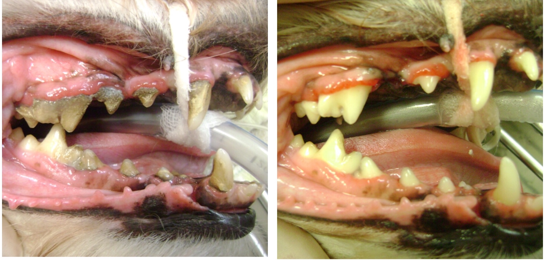
Cleaning begins by removing large pieces of tartar by hand scaling both above and below the gum line. Ultrasonic scaling will be used to remove any material that was left behind. Once again the mouth is visibly inspected for any remaining tartar, and a probe is run over the tooth to “feel” for any irregularities caused by tartar left on the tooth (VetMed Team).
After scaling is complete the teeth are then polished. Scaling can create micro abrasions on the tooth’s surface. If these abrasions are not smoothed out, they will provide a foothold for plaque and tartar formation. Finally the mouth is rinsed with water, to flush out any debris, and air dried. A fluoride treatment is applied to the teeth to strengthen the enamel.

Home Care
The true definition of prophylaxis is “the prevention of disease or control of its possible spread (Farlex).” A dental cleaning is a treatment of a disease process, whereas true prophylaxis is done at home. Home therapy is targeted at removing or lowering the amount of bacteria in the mouth. If home care is not instituted by three months after a dental cleaning then gingivitis scores are equivalent to those recorded prior to dental cleaning (Gorrel, 2004).

Brushing teeth is the gold standard for at home dental care; however, there are many options available. Products range from traditional tooth paste and tooth brush, to toys, treats, chews, wipes, and sprays. This allows us to design the home care plan that fits your pet’s needs and your lifestyle as well. Home care is a large subject matter and will be the focus of a later blog.

Conclusion
True dental prophylaxis encompasses both the professional dental cleaning under anesthesia coupled with home care. Dental cleanings that are incomplete or focus on the visible tooth anatomy can regress quickly. To answer my opening question; No, we cannot just simply scrape that off his teeth.

Works Cited
Farlex. (n.d.). The Free Dictionary. Retrieved February 1, 2013, from http://www.thefreedictionary.com/prophylaxis
Gorrel, C. (2004). Veterinary Denistry for the General Practitioner. Edinburgh, London, NY, Oxford, Philadelphia,St. Louis, Sydney, Toronoto: Saunders.
Iams Partners for Health. (2003). Spotlight on Dentistry: More than meets the eye.
VetMed Team. (n.d.). Companion Animal Denistry 101. Retrieved January 20, 2013, from www.vetmedlearn.com
VetMed Team. (n.d.). Companion Animal Dentistry 102. Retrieved January 20, 2013, from www.vetmedlearn.com