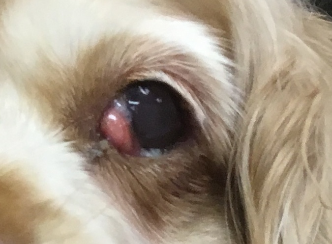
Category

Medical Conditions

Cherry Eye

By Medical Conditions No Comments

Cherry Eye

Cherry eye is the common name for a prolapsed gland of the third eyelid.  The eyes contain a series of glands with the purpose of lubricating the eye.  This continual flushing keeps the eye clean by rinsing off surface debris.  One of the glands that helps keep the eye moist is the third eyelid gland (nictitating membrane).  This gland sits below the eye and points towards the nose. It produces up to 50% of the eye’s tears.

The third eyelid gland is held in place by fibrous connective tissue.  If the fibrous attachment is weak it predisposes the gland to prolapse, or simply, fall out of place.  When the gland prolapses a red mass is seen protruding from the inner corner of the eye, hence the name cherry eye.  One or both eyes may prolapse, and if it occurs in one eye the unaffected eye may prolapse later.  In addition to the gland protruding, dogs may experience an overflow of tears onto the face, red eye, and/or eyelid spasms.

Certain breeds more prone to cherry eye include Beagles, English Bulldogs, Cocker Spaniels, and other purebred or mixed dogs with droopy eyes, such as hounds.  If congenital in nature, prolapse usually occurs anywhere from 6 months to 2 years of age in dogs.  The cause of cherry eyes in dogs over 2 years of age, may include trauma or even more serious – tumors that may be pushing the gland out of place.

Historically, the treatment for cherry eye involved surgically removing the prolapsed glands.  This procedure was used before the full significance of the gland was realized.  When surgically removed, dogs may be predisposed to develop keratoconjunctivitis sicca (dry eye) later in life.   Today veterinarians prefer surgical correction. This procedure re-positions the gland into its normal position and is sutured in place.  After surgery a topical anti-inflammatory and elizabethan collar are sent home.  The collar prevents the patient from rubbing or scratching the eye, which can disrupt the sutures causing prolapse to reoccur and/or corneal ulcers.

Prognosis for dogs undergoing surgery is good, with only a 5-20% chance of relapse.  Cherry eye can be scary in appearance, especially if you have never seen it, but it is not a medical emergency.  Schedule an appointment with your veterinarian within a day or two.


Hot Spots

By Medical Conditions No Comments

Hot Spots

The hot, humid summer months can bring fun in the sun for our pets, but they can also bring seasonal medical concerns.  During this time of year, we see an increase in the number of pets suffering from hot spots.  Hot spots are patches of red, moist, itchy, painful, and often hairless patches of skin.  Hot spots (acute moist dermatitis) are skin infections affecting the superficial layer of skin. They generally heal rapidly and often heal without scarring.  Hot spots can occur very fast and are suspected to be caused by self trauma.  One example of self-trauma is a hot spot below an infected ear.  The pet has scratched around the ear, damaging the protective layer, and allowing an infection to form.

While hot spots can occur at any time they are most commonly seen during the hot, moist summer months through early fall.  Breeds most affected by hot spots are heavy coated, water loving breeds such as the Golden Retriever, Labrador Retriever, Newfoundland, Saint Bernard, and German Shepherd Dog.

Diagnosis is based on clinical signs and history described by owners.  A thorough exam is done to rule out underlying causes such as skin parasites (fleas, ticks, etc.), allergies, or infections.  We begin treatment right away by clipping hair both in and around the lesion.  For large or especially painful regions sedation may be necessary.  After the hair is removed the technicians will gently clean the lesion with wound cleanser.

Treatment at home depends on severity of the lesion/s and location.  Elizabethan collars, (cone collars), are often sent home to prevent licking or chewing which can further traumatize the lesion.  Your veterinarian may send home oral antibiotics, and/or steroids to treat the skin infection and inflammation.  Cleaning of the lesions daily is recommended to speed the healing process – often followed by a topical medication such as a cream, ointment, or powder.  It is important to wash your hands after cleaning and treating a hot spot to prevent the spread of bacteria.

Pets suffering from hot spots recover quickly with treatment.  However relapse is common depending on the underlying cause.  Hot spots are not a medical emergency, but they should be addressed by a veterinarian quickly.  The lesions are painful and advance quickly with persistent scratching and /or chewing.

Luxating Patella

By Medical Conditions No Comments


Luxating Patella

Our smaller patients frequently suffer from luxating patellas, a dislocated knee.  This condition is common in many small and toy breed dogs, most notably the Toy Poodle and Yorkshire Terrier.  Though not as common, cats can also suffer from luxating patellas.

The knee joint is comprised of four bones, the femur, patella, tibia, and fibula.  Just like a human knee, large tendons hold the patella in place.  The patella sits in the patellar groove on the front of the femur.  In a normal knee, the patella moves up and down in its groove as the knee straightens and extends.

In a knee with patellar luxation, the patella dislocates out of its groove to the inside or outside of the groove.  About 75% of patients have patellas that move to the inside of the leg.  Over time this movement causes wear to the cartilage that covers the knee joint.  This can cause pain and degenerative joint disease.

The most common sign of luxating patellas is intermittent rear limb lameness ranging from a limp to non-weight bearing.  Diagnosis can be made by feeling the movement of the patella or visually on radiographs.  Once diagnosed, this condition is classified into one of four grades.

  • Grade I patellar luxation rarely shows clinical signs. Diagnosis is most often made during yearly preventative health exams by manually moving the patellar out of position.  However it will spontaneously correct itself.
  • Grade II may appear as a skip in the pet’s gait. The patella will move out of place as they are running then slip quickly back into place.  When diagnosing, the patella will move in and out on its own as the leg is bent and straightened.
  • Grade III and IV are uncomfortable for the pets and affect their gait significantly. Pets may limp or not place any weight on the limb.  When diagnosing a grade III the patella will move out of place when the leg is bent and straightened and needs to be manually replaced.  With grade IV the patella will move out of place and cannot be replaced manually.

Treatment is based on the grade of luxation.  Grade I luxation rarely requires treatment.  Grade II are evaluated on a case by case basis, and may respond well to anti-inflammatory medications.  Grade III and IV generally require surgical correction.  There are two procedures to correct a luxating patella.  The first involves deepening the groove the patella sits in.  The second repositions a part of the tibia which straightens the leg, reducing the tension pulling the patella out of place.

Prognosis is usually good for all pets suffering from patellar luxation.  Changes to exercise routines, medications, and surgical treatments can help alleviate any discomfort a pet may suffer from this condition.


Canine Influenza

By Medical Conditions No Comments

Canine Influenza


Canine Influenza, or dog flu, is a highly contagious virus that affects dogs as well as cats. Canine Influenza is highly contagious and easily spread from infected dogs to other dogs. Almost every dog exposed to the virus will become infected. Recently, both strains (H3N8 and H3N2) have been (and still are) being diagnosed in Macomb County.

The virus is transmitted by either direct contact, in the air (barking, coughing, sneezing), contaminated objects, and by people moving from infected to uninfected dogs. The virus can live on surfaces for 48 hours, clothing for 24 hours, and hands for 12 hours.

Symptoms mimic kennel cough but the clinical signs of the cough persist for 10 to 21 days despite treatment.  Affected dogs may have a soft, moist cough, nasal or eye discharge, sneezing, lethargy, or decreased appetite.  Many dogs will spike a fever up to 105 degrees. Some, more severely affected, may exhibit signs of pneumonia.

What can you do to keep your dog safe?  Vaccinate with the bivalent H3N8/H3N2 vaccine.  This vaccine requires a booster 3 weeks.  Until then…

  • Avoid frequent dog areas such as dog parks, pet day care, grooming facilities, kennels, dog-friendly stores, and communal water bowls.
  • Wash your dog’s toys, bowls, and bedding regularly.
  • When in contact with other dogs, even if they don’t appear sick, wash your hands and change your clothes before handling your own pet.
  • Avoid contact with sick or possibly exposed dogs as the virus can persist on clothing and other surfaces for at least 24 hours.


Call Veterinary General for more information and to schedule your dog’s vaccine today!  586-992-3810


UNDERSTANDING BLOAT

By Medical Conditions No Comments

Bloat, a Flip Flop Stomach

            Bloat is a condition that affects the GI system, mostly the stomach, but other body systems including the circulatory system can be affected. Food and water enter the stomach through the esophagus and exits into the small intestine after mixing with digestive enzymes.  The stomach is very similar to a balloon; both have a remarkable ability to expand many times their normal size.  A rapidly expanding stomach can cause bloat, which occurs in two forms gastric dilation and gastric dilation-volvulus (GDV).

In gastric dilation, commonly referred to as food bloat, the stomach can expand many times its normal size displacing other organs and blood vessels as it expands. This stage of bloat is very uncomfortable for the dog, but can become dangerous if the stomach rotates.  At this stage the affected dog will still need to see their veterinarian and stay as calm as possible.

In gastric dilation-volvulus the large, heavy stomach can rotate over itself, similar to twisting a balloon before knotting it.  Food cannot enter or leave the stomach, however the stomach continues to expand as gas is produced and digestive enzymes are secreted.  The rotation and expansion can cut off blood flow to the stomach itself and have a detrimental affect on blood flow returning to the heart.  These dogs can have arrhythmias, or abnormal heart rhythms.  GDV is life threatening and can kill a dog within hours.

Breeds that are most likely to be affected are deep-chested, large breed dogs, including Great Danes, Greyhounds, St. Bernards, and Weimaraners (to name a few).  While large breeds are more prone to bloating, any dog, no matter their size can suffer from bloat.  Other risk factors include dogs that only eat one meal a day, have a history of bloat, eat very quickly, and/or dogs that exercise after a meal.

Symptoms of GDV can progress very rapidly.  Dogs may show signs of nausea include drooling, they may also retch but not bring anything up.  Dogs can have abdominal distension and pain which can lead to refusal to eat or drink, pacing, and restlessness.  Upon physical exam by a veterinarian a distended abdomen can lead to a diagnosis of bloat, however radiographs are needed for further diagnosis of bloat with or without torsion of the stomach.

Once a veterinarian has confirmed diagnosis a treatment plan can be made.  Many factors such as type of bloat, medical history, breed, and state of shock will determine which treatment option to take.  Most commonly bloat is treated surgically or by decompressing the stomach, however depending on the severity other options may be available.  Statistically 75% of dogs that bloat will bloat again without surgery.  Once stable most dogs undergo a gastropexy procedure, a surgery to tack the stomach to the abdominal wall so it cannot rotate over itself again.

If you feel your dog may be suffering from bloat take them to your veterinarian immediately.  Bloat can go from serious to life threatening very quickly and prompt care is vital.  You can decrease your pet’s risk of bloat by feeding smaller meals instead of a single large meal, elevating the food bowl, avoiding exercise after eating, and encouraging your dog to eat slower by adding water, using a specially designed bowl, or interactive food toys.  High risk breeds may elect to have a gastropexy performed at the same time as their spay or neuter as a preventative.

Veterinary General is located in Shelby Township, Michigan. We offer traditional and alternative therapies such as Acupuncture, Chinese Herbals, and Cold Laser Therapy. More information can be found at www.veterinarygeneral.com.